Horses are a vital part of our sport. No horse, no riding! When you participate in a sport, it’s inevitable that sometimes, your equipment needs repair. Maybe your tennis racket gets a hole and needs to be fixed up, or maybe your swimming goggles strap broke, so you have to buy another pair.
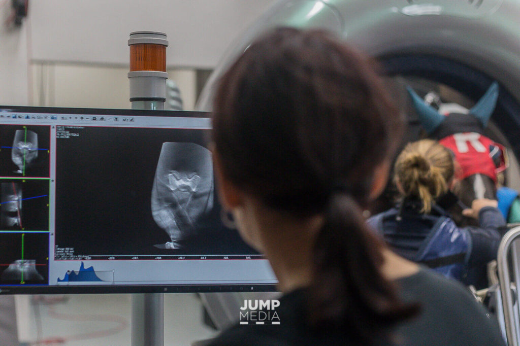 Reviewing a CT scan at Palm Beach Equine Clinic. Photo by Jump Media.
Horses, however, are our live partners. We can’t just buy a new leg and strap it on if our horse comes up sore one day. Also, as living, breathing sporting partners, it can be difficult to diagnose where exactly an issue is coming from if our horse needs “repair.” That’s where all of the advanced imaging technologies that exist today for lameness evaluations can help.
Before learning about these technologies, I was really only familiar with MRI and endoscopy procedures — but there’s so much more. Let’s dig in.
 Nuclear Scintigraphy (Bone Scan)
Nuclear scintigraphy begins with the injection of a radioactive isotope called Technetium 99 that is bound to a phosphate analogue. The isotope – phosphate molecule attaches to the mineral matrix of the bone in areas where bone is active. A gamma camera is then used to capture images of the skeletal anatomy. Points that “light up” on the image indicate increased metabolic activity as a possible site of injury.
Photo by Palm Beach Equine Clinic.
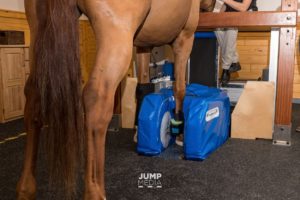 Standing Magnetic Resonance Imaging (MRI)
The standing MRI produces highly detailed, cross-sectional images of bone and soft tissue in multiple different planes to fully image a desired region. The standing MRI requires sedation (not anesthesia) and is best used to further define a specific area that has already been pinpointed as the origin of lameness.
Photo by Jump Media.
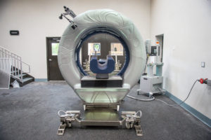
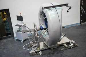 Computed Tomography (CT)
Similar to its use in humans, CT allows veterinarians the unique opportunity to conveniently explore areas of a horse’s body that were previously inaccessible. The machine produces 360 degree images of a horse’s neck, spine, and head and can be conducted while a horse is standing and under only light sedation.
Photo by Erin Gilmore.
 Endoscopy
An endoscope is an instrument that allows veterinarians to look inside the body by being inserted through a natural opening or incision. A tiny camera on the instrument allows an in-depth view of internal structures such as the upper and lower respiratory track, gastrointestinal and urinary tracts, as well as the cervix and uterus of mares.
Photo by Palm Beach Equine Clinic.
 Digital Radiography (X-Ray)
Used routinely, radiographs are traditional x-rays that are made available within seconds for digital viewing and evaluation.
Photo by Jump Media.
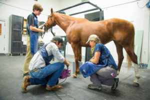
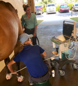 Ultrasonography
An ultrasound machine generates high-frequency sound waves, which echo an image back to the machine where bone appears white, fluid appears black, and all other structures are on a grayscale. An ultrasound is non-invasive, usually does not require sedation, does not use radiation or require injecting radioactive isotopes, and provides real-time images.
Photo by Jump Media.
Palm Beach Equine Clinic radiologist Dr. Sarah Puchalski explains, “These tools can give a definitive diagnosis, and that saves time and money in the long run. For example, if a horse goes lame and it gets seen and treated empirically, which is a diagnosis based on likely problems through common diagnostic procedures, it either stays sound or it becomes lame again or even non-functional in three to six months. This method sets back the commencement of the appropriate therapy. What these modalities do is allow the horse to be treated early and correctly. Otherwise, you may not be treating the correct issue, and the horse could end up lame again very soon.”
Ultimately, if the lameness is diagnosed exactly for what it is, the horse can be treated correctly from the start and be well again sooner, rather than regressing.
Have you used any of these types of diagnostic technologies before for your horse? It’s amazing to see how how far veterinary medicine has come to help our horses get well sooner.
Diagnostic description words by Beth Lawler.